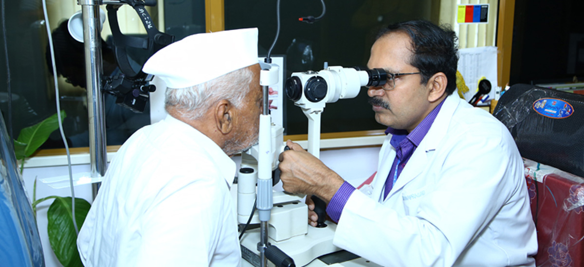
Advanced Diagnosis and Treatment for Glaucoma Patients

At M M Joshi Eye Institute, our Glaucoma Clinic offers a complete range of diagnostic and therapeutic services for patients with glaucoma. We specialize in early detection, ongoing management, and advanced treatment options to help prevent vision loss caused by this silent, progressive disease.
State-of-the-Art Diagnostic Equipment
We use cutting-edge instruments to provide the most accurate and thorough glaucoma evaluations, ensuring our patients receive the best care possible. Our diagnostic tools include:
Goldman Applanation Tonometer, Perkins Tonometer, iCare No Anesthesia Tonometer, and Non-Contact Tonometer for precise intraocular pressure (IOP) measurements
Humphrey Perimeter for visual field examination to detect visual field loss.
Topcon Fundus Photography for detailed optic disc documentation with color and red-free images.
Optical Coherence Tomography (OCT) for optic disc imaging and retinal nerve fiber layer analysis.
What is Glaucoma?
Glaucoma is one of the leading causes of irreversible blindness, particularly affecting older adults. It occurs when the optic nerve, which carries visual information from the eye to the brain, becomes damaged due to increased pressure inside the eye. Over time, this damage can cause significant vision loss if left untreated.
Why is Glaucoma Dangerous?
Often referred to as the “silent thief of sight,” glaucoma is painless, has no early symptoms, and can cause irreversible vision loss. In fact, nearly half of those with glaucoma are unaware they have it. Early detection and treatment are critical to prevent blindness.
Types of Glaucoma
Chronic Open-Angle Glaucoma: The most common form of glaucoma, it progresses slowly and painlessly. Vision loss occurs gradually, often without noticeable symptoms.
Angle-Closure Glaucoma: An acute form that occurs when the drainage channels in the eye suddenly become blocked, causing a rapid increase in pressure. Symptoms include severe eye pain, blurred vision, nausea, vomiting, and seeing rainbow halos around lights. This is a medical emergency that requires immediate attention.
Glaucoma Detection and Diagnosis
Early detection through regular eye exams is crucial for managing glaucoma. Our thorough and painless diagnostic tests include:
Tonometry: Measuring intraocular pressure.
Gonioscopy: Inspecting the drainage angle of the eye.
Ophthalmoscopy: Evaluating optic nerve damage.
Perimetry: Testing the visual field of each eye.
Glaucoma Treatment Options
Though glaucoma damage is irreversible, treatment can slow or prevent further vision loss. Treatment options include:
- Eye Drops: These reduce intraocular pressure by either decreasing fluid production or improving drainage.
- Tablets: Sometimes prescribed in combination with eye drops to further lower eye pressure. Long-term use can occasionally cause side effects such as tingling in the fingers or toes and kidney stones.
- For open-angle glaucoma, lasers can help enlarge the drainage area.
- For angle-closure glaucoma, lasers create a hole in the iris to improve fluid drainage.
- In advanced glaucoma cases, lasers are used to reduce fluid production in the eye.
- Trabeculectomy: Creating a new drainage channel for the eye
- Glaucoma Drainage Device: A tube may be inserted to help drain fluid in resistant cases.
Specialized Instruments in the Glaucoma Clinic
Apart from routine evaluation tools, we have specialized equipment to manage complex cases:
Tonopen and Perkins Handheld Applanation Tonometer for accurate pressure readings.
Koeppe Direct Gonioscope: For pediatric angle assessment.
Anterior Segment OCT and Ultrasound Biomicroscopy (UBM): For detailed imaging of the eye’s structures.
Fundus Photography Unit: For stereoscopic disc photography.
Outpatient and Diagnostic Services
We offer comprehensive outpatient services for glaucoma patients, ensuring regular monitoring and timely interventions:
Routine screening, including tonometry, gonioscopy, and optic disc evaluation.
Visual field analysis using the Humphrey Visual Field Analyzer.
Disc and gonioscopic photography when needed.
Management of pediatric glaucomas and complex cases referred from other departments.
Educational and Research Activities
We are committed to advancing glaucoma care through Continuing Medical Education (CME) programs and anti-glaucoma drug trials. Our research-driven approach ensures that patients receive the latest, most effective treatments available.
Our Glaucoma Service Experts
Why Choose M M Joshi Eye Institute for Glaucoma Care?
Schedule Your Glaucoma Screening Today!
If you are at risk of glaucoma or experiencing any symptoms, book an appointment with our glaucoma specialists for a comprehensive evaluation.
FAQs
Glaucoma is a disease of the optic nerve, a structure that connects the eye to the brain. The nerve is composed of 1.2 million fibers that originate in the retina. If the optic nerve is damaged from any cause, visual signals from the eye cannot reach the brain, leading to vision loss.
In glaucoma, the optic nerve fibers degenerate over time. The nerve then remodels, or changes shape, as the tissue is lost – this process is called “cupping.” These nerve changes occur in characteristic patterns which are distinct from other optic nerve diseases and can be identified during a dilated eye exam. Uncontrolled glaucoma is a progressive disease characterized by increased cupping over time.
In general, the fibers that are responsible for peripheral vision are affected first, and those maintaining central vision are affected late in the disease. As a result, most people with glaucoma will maintain vision straight ahead in at least one eye. Unfortunately, people may not notice changes in their peripheral vision in their everyday life, and so they may be unaware of a problem until they already have advanced damage. The optic nerve is an extension of the brain – like the brain and spinal cord, the optic nerve does not regenerate. As a result, any vision lost secondary to optic nerve damage is permanent. This is why early detection and treatment are absolutely essential for preventing blindness from glaucoma.
This is a complicated question which many researchers, are trying to understand. From ongoing research, we know that genetics, eye pressure and blood flow are all involved. While high eye pressure (over 21 mm Hg) is commonly associated with glaucoma, it is not part of the definition of glaucoma. A person can develop glaucoma at normal eye pressures (13 to 20 mm Hg). For this reason, optic nerve examination is the key to diagnosis.
Glaucoma can be diagnosed with a complete eye examination. This includes measuring vision and eye pressure and having a dilated eye examination to properly evaluate the optic nerve. If any of these parameters are abnormal (for example, high eye pressure or a suspicious appearing optic nerve), further testing will be done.
Glaucoma is a chronic disease. Using your eye drops faithfully and having regular examinations to monitor the eye pressure and optic nerve are the two most important parts of glaucoma care. Once the eye pressure is controlled, it will be monitored every 3-6 months. This is essential because pressure can increase over time despite medications. Since even moderately high eye pressure rarely causes pain, you will not be able to tell if your pressure is elevated and need your doctor to check it on a regular basis. Be prepared for regular checks of your optic nerve visual field. While we know visual fields can be hard to take, they are particularly sensitive at detecting changes if you already have an abnormal visual field. Glaucoma is not about a pressure. While we take into account a lot of parameters to pick a general target for your pressure, everyone is different and each nerve reacts differently to pressure. Treatment is a dynamic process, and we continually reassess our goals based on how the eye is responding to certain levels of pressure. If your nerve or field changes, we will adjust our treatment to lower your pressure even further using surgery (trabeculectomy).

